Three zone streaking method

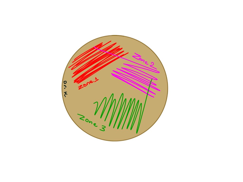
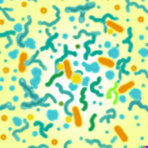
3-zone streaking pattern

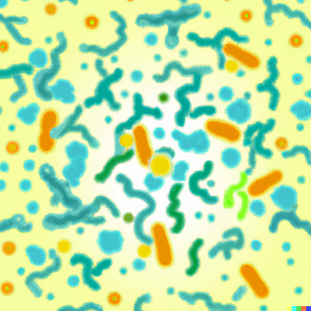
After incubation of 3-zone streaked plate

The three-zone streaking method is a laboratory technique used to isolate and identify bacterial strains. It involves using a sterile inoculating loop to streak a culture of bacteria onto a Petri dish containing agar. The agar is a solid, nutrient-rich growth medium that allows the bacteria to multiply and form colonies.

The streaking process involves making three separate streaks on the Petri dish, each in a different area. The first streak is made in the center of the dish, the second streak is made in the middle section of the dish, and the third streak is made in the outer section of the dish.

As the bacteria grow, they form colonies on the agar. The colonies that form in the center of the dish are the most densely populated and are called the confluent zone. The colonies that form in the middle section of the dish are less densely populated and are called the intermediate zone. The colonies that form in the outer section of the dish are the least densely populated and are called the marginal zone.

The three-zone streaking method allows the bacteria to be isolated and identified based on the size and appearance of the colonies they form. For example, a large, circular colony with a smooth, shiny surface may indicate the presence of Staphylococcus aureus, while a small, circular colony with a rough, granular surface may indicate the presence of Escherichia coli.

Classification of viruses

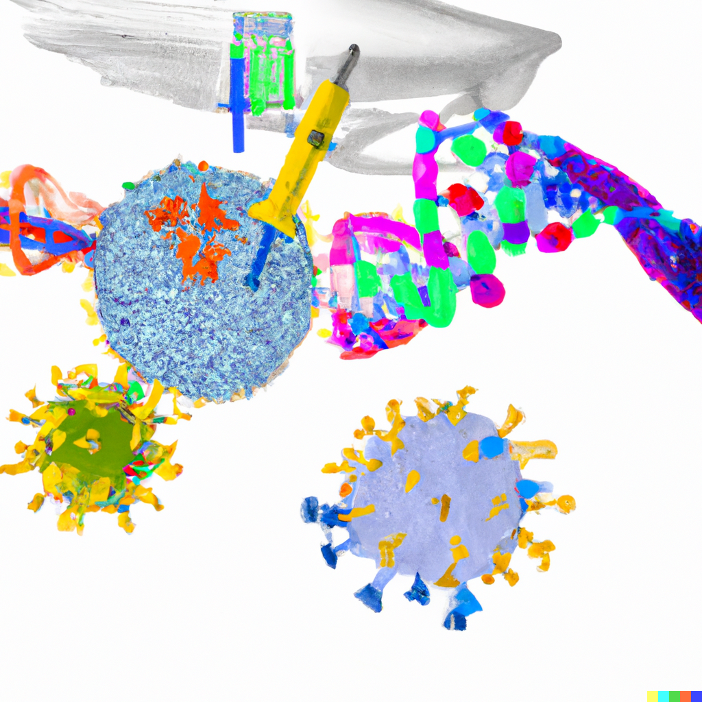
Viruses are small infectious agents that can replicate only inside the living cells of an organism. They are typically classified based on their shape, size, and the type of disease they cause.

The most common way to classify viruses is by their shape. There are four main shapes of viruses: 

Spherical or virion viruses: These viruses have a round shape, like a ball. They are often the smallest type of virus and can range in size from 20 to 400 nanometers in diameter. Examples of spherical viruses include the influenza virus and the common cold virus.

Helical viruses: These viruses have a spiral shape, like a spring or a coil. They are typically longer than virions and can range in size from 100 to 1,000 nanometers in diameter. Examples of helical viruses include the tobacco mosaic virus and the hepatitis C virus.

Icosahedral viruses: These viruses have a shape like a soccer ball, with 20 identical triangular faces. They are the most common type of virus and can range in size from 25 to 250 nanometers in diameter. Examples of icosahedral viruses include the human papillomavirus and the poliovirus.

Complex viruses: These viruses have a more irregular shape, with multiple structures or components. They can range in size from 200 to 1,000 nanometers in diameter. Examples of complex viruses include the HIV virus and the adenovirus.Another way to classify viruses is by their size. Viruses range in size from 20 nanometers to several micrometers in diameter. The smallest viruses, such as the tobacco mosaic virus, are only 20 nanometers in diameter, while the largest viruses, such as the poxviruses, can be up to 500 nanometers in diameter.

Viruses can also be classified based on the type of disease they cause. The most common types of viral diseases include:

Respiratory infections: These infections affect the respiratory system, including the nose, throat, and lungs. Examples of respiratory viruses include the influenza virus, the common cold virus, and the coronavirus.

Gastrointestinal infections: These infections affect the digestive system, including the stomach and intestines. Examples of gastrointestinal viruses include the norovirus, the rotavirus, and the hepatitis A virus.

Sexually transmitted infections: These infections are spread through sexual contact and can affect the genitals, anus, or mouth. Examples of sexually transmitted viruses include the HIV virus, the herpes simplex virus, and the human papillomavirus.

Other viral diseases: These diseases can affect various other systems in the body, such as the liver (hepatitis), the brain (encephalitis), and the skin (chickenpox). Examples of viruses that cause these diseases include the hepatitis B virus, the West Nile virus, and the varicella-zoster virus.

There are also several sub-classifications of viruses based on their specific characteristics. For example, some viruses are classified as enveloped viruses because they have a protective layer of proteins and lipids surrounding their genetic material. Other viruses are classified as non-enveloped viruses because they do not have this protective layer.

Additionally, some viruses are classified as DNA viruses because they contain DNA as their genetic material, while others are classified as RNA viruses because they contain RNA. There are also retroviruses, which are a type of RNA virus that can convert their RNA into DNA and integrate it into the host cell’s genome.


I am profound

A single cell, so small and round

In a world of microbes, I am profound

I reproduce quickly, with no need for mates

I adapt and evolve, it’s simply my fate

I can be helpful, or cause harm and pain

I exist in every place, from soil to the vein

I am a bacterium, and this is my story

A never-ending journey, without any worry

I may be tiny, but I am mighty and strong

I have survived for ages, and I will live on

Through the ages I’ve been, and I will always be

A tiny, yet powerful, part of this world’s history.

What is somatic mutation theory of the origin of cancer?

The somatic mutation theory of cancer is a proposed mechanism for the development of cancer in mammals. It suggests that cancer arises from mutations in the genes of somatic (non-reproductive) cells.

According to the somatic mutation theory of cancer, the development of cancer is a multistep process that involves the accumulation of mutations in the genes of somatic cells. These mutations can arise from a variety of sources, including environmental factors (such as exposure to radiation or chemicals), errors during DNA replication, and inherited genetic predispositions.

The somatic mutations that occur in cancer cells can affect a variety of genes that are involved in cell growth and division. For example, mutations in genes that encode for proteins that regulate cell division, such as the p53 gene, can cause uncontrolled cell growth and proliferation. Mutations in genes that encode for proteins that repair damaged DNA, such as the BRCA1 gene, can also lead to the accumulation of additional mutations in cancer cells.

The somatic mutation theory of cancer is supported by evidence from laboratory experiments, where cancer cells have been shown to have a higher number of mutations than normal cells. It is also supported by observations of cancer in humans, where different types of cancer have been shown to have different patterns of genetic mutations.

However, the somatic mutation theory of cancer is not without its critics. Some researchers argue that other mechanisms, such as epigenetic changes or viral infections, may also play a role in the development of cancer. Additionally, the somatic mutation theory does not explain why some people are more susceptible to developing cancer than others, or why some cancer treatments are more effective than others.

Despite these criticisms, the somatic mutation theory of cancer remains a valuable framework for understanding the development of cancer in mammals. By studying the mechanisms of somatic mutation and the genes that are affected by these mutations, researchers can develop more effective treatments for cancer and improve the prognosis for cancer patients.

Eukaryotic cell cycle

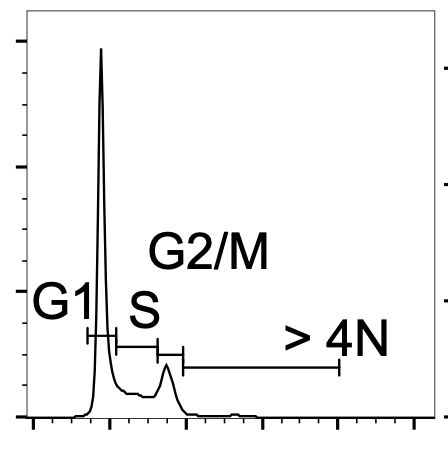
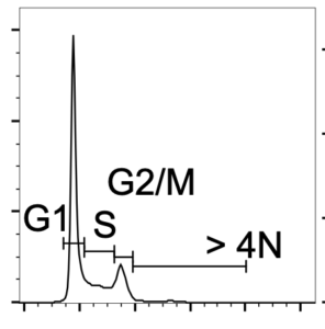
Figure: Cell cycle map of eukaryotic cells. The X-axis represents DNA content, and Y-axis represents the number of cells. 

The cell cycle is the process by which a cell grows and divides into two daughter cells. It is a complex and tightly regulated process that is essential for the growth and development of all living organisms.

The cell cycle can be divided into two main phases: interphase and cell division. Interphase is further divided into three stages: G1, S, and G2. Cell division is divided into two stages: mitosis and cytokinesis.

During interphase, the cell grows and performs its normal functions. In the G1 stage, the cell grows and carries out its usual functions. In the S stage, the cell’s DNA is replicated, creating two identical copies of the genome. In the G2 stage, the cell prepares for division.

During mitosis, the cell’s nuclear material is divided into two equal sets, each of which will be inherited by one of the daughter cells. This is accomplished through a series of highly coordinated events.

First, the nuclear envelope, which surrounds the nucleus, breaks down and the chromosomes that carry the cell’s genetic material condense and become visible under a microscope. The chromosomes are then pulled to opposite poles of the cell by the mitotic spindle, a structure made up of microtubules.

Next, the cell’s cytoplasm, which contains all of the cell’s organelles, divides into two equal parts, each of which will be inherited by one of the daughter cells. This is accomplished through a process called cytokinesis.

In animal cells, cytokinesis is accomplished through the formation of a cleavage furrow, which is a shallow indentation that forms in the cell’s plasma membrane. As the cleavage furrow deepens, the cell’s cytoplasm is divided into two daughter cells.

In plant cells, cytokinesis is accomplished through the formation of a cell plate, which is a structure that forms in the middle of the cell. The cell plate then grows outward, eventually forming a new cell wall that divides the cell into two daughter cells.

After cytokinesis, the cell cycle is complete and the two daughter cells are ready to begin their own cell cycles.

Overall, the cell cycle is a complex and highly regulated process that is essential for the growth and development of all living organisms. It allows cells to grow, divide, and produce new cells that are genetically identical to the parent cell. Understanding the stages of the cell cycle is important for many areas of biology, including cancer research and the development of new treatments for diseases.

Brewing booze 🍺

Once upon a time, in a land far, far away, fermentation was discovered by a group of merry microorganisms who were feeling a bit peckish. They stumbled upon a delicious bowl of carbohydrates and, being the clever critters they were, they figured out that they could turn those carbs into alcohol or acid. And thus, fermentation was born!

From that day forth, fermentation became the go-to method for producing all sorts of tasty treats. The ancient Egyptians used it to make bread and boozy beverages, while the Chinese used it to create soy sauce, rice wine, and pickled veggies. The Europeans, being the beer-loving bunch that they are, used fermentation to brew up batches of their favorite frothy beverage.

But fermentation wasn’t just for fun and games. It was also used for practical purposes, like preserving food and making medicine. In the Middle Ages, sauerkraut was all the rage, and fermented drinks were believed to have magical healing powers.

Fast forward to today, and fermentation is still going strong. We now have fancy gadgets like microscopes and fancy-pants scientists who have figured out all the nitty-gritty details of the fermentation process. So pop open a cold one and raise a glass to fermentation – the original party trick of the microbe world!

In more recent history, the science of fermentation has advanced significantly. The development of the microscope in the 17th century allowed scientists to study microorganisms up close and personal, and the identification of yeast as the primary agent of fermentation in the 19th century paved the way for the modern understanding of the process. Today, fermentation is used in the production of a wide range of products, from food and drink to pharmaceuticals and biofuels.

No doubt, the history of fermentation is a long and fascinating one. Full of twists and turns and the occasional belch. It’s a tale of clever microorganisms, boisterous brewmasters, and countless cultures coming together to create something truly special. So here’s to fermentation – may it continue to bring joy, flavor, and a little bit of gas to our lives for many centuries to come!

On scientific method

The scientific method is a systematic approach to conducting research and solving problems. It is a process that is used by scientists to explore the natural world, to test hypotheses, and to develop theories.

The scientific method typically begins with the observation of a phenomenon or problem. This observation leads to the formation of a hypothesis, which is an educated guess or prediction about the phenomenon or problem. The hypothesis is then tested through experimentation or observation, and the results of the experiment are analyzed to determine whether the hypothesis is supported or rejected.

If the hypothesis is supported, it may be accepted as a scientific theory. A scientific theory is a well-supported and widely accepted explanation of a phenomenon or problem. However, even a widely accepted theory is always subject to further testing and refinement, as new evidence or observations may lead to new hypotheses and theories.

The scientific method is a critical tool for advancing our understanding of the world around us. It is a systematic and rigorous approach that helps to ensure that scientific research is objective and accurate. It also allows scientists to build upon the work of others, to develop new theories and technologies, and to make progress in addressing some of the most pressing challenges facing humanity.

The scientific method is not always a straightforward or linear process. It can involve many different steps, and it can require significant time, effort, and resources to complete. However, the scientific method is a critical tool for advancing our understanding of the world and for solving some of the most complex and important problems that we face.

How does RNA interference (RNAi) work?

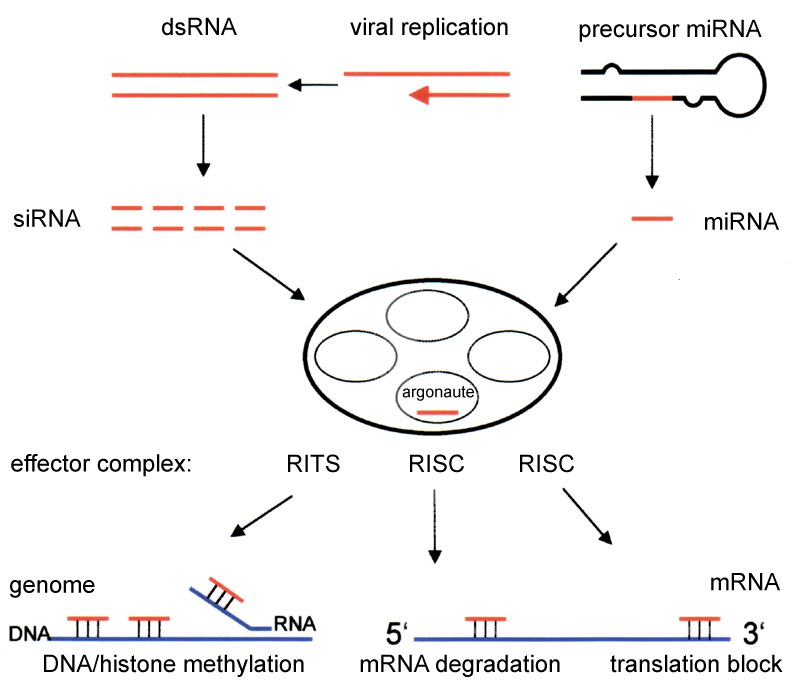
Picture source: Wikipedia

RNA interference (RNAi) is a mechanism that cells use to regulate gene expression. Small interfering RNAs (siRNAs) are a type of RNA molecule that plays a key role in the RNAi process. SiRNAs are short, double-stranded RNA molecules that are typically 21-23 nucleotides in length.

When a cell is exposed to siRNAs, they are recognized by an enzyme called Dicer, which cuts the siRNA molecules into short pieces called siRNA duplexes. The siRNA duplexes then bind to a protein complex called the RNA-induced silencing complex (RISC), which uses one of the strands of the siRNA duplex as a guide to find and bind to complementary RNA molecules.

Once the RISC complex has bound to a complementary RNA molecule, it uses an enzyme called an RNase to degrade the RNA molecule, preventing it from being translated into a protein. This process is called gene knockdown.

Gene knockdown is often used in research to study the effects of reducing the expression of a specific gene. By using siRNAs to knock down the expression of a gene, researchers can study the effects of reducing the levels of the corresponding protein. This can provide important insights into the function of that gene and the role of the corresponding protein in cell function.

One of the key advantages of using siRNAs for gene knockdown is that they can be designed to target specific genes. This allows researchers to study the effects of reducing the expression of a specific gene, without affecting the expression of other genes.

Additionally, siRNAs can be delivered to cells using a variety of methods, including transfection with vectors such as plasmids or viruses, or by direct injection into cells or tissues. This makes it possible to study the effects of gene knockdown in a wide range of cell types and organisms.

Overall, siRNA-mediated gene knockdown is a powerful tool for studying gene function. By using siRNAs to reduce the expression of specific genes, researchers can gain important insights into the role of those genes in cell function and disease.

Can Molecular Biology Support The Theory Of Evolution

Hedgehog model for studying evolution. Picture source: Wikipedia.

The theory of evolution is the scientific explanation for the diversity of life on Earth, and is supported by a wealth of evidence from many different fields of study, including molecular biology. Molecular biology is the study of the structure and function of macromolecules, such as nucleic acids and proteins, which are essential to life. By studying the molecular mechanisms of life, molecular biologists have been able to provide important insights into the processes of evolution.

One of the key ways in which molecular biology supports the theory of evolution is through the study of the genetic code. The genetic code is the sequence of nucleotides in DNA and RNA molecules, which carries the information necessary for the synthesis of proteins. By studying the genetic code, molecular biologists have been able to demonstrate that all living organisms share a common ancestry, and that the genetic code is highly conserved across different species.

Another way in which molecular biology supports the theory of evolution is through the study of molecular evolution. Molecular evolution is the study of the changes in the sequences of nucleotides and amino acids over time, and is a key mechanism by which evolution occurs. By studying the molecular changes that have occurred over time, molecular biologists have been able to demonstrate that different species are related to one another, and that the evolutionary history of life on Earth can be reconstructed.

Molecular biology has also contributed to the understanding of natural selection, which is the process by which certain traits become more or less common in a population over time. Molecular biologists have been able to demonstrate that natural selection can act at the molecular level, and that changes in the sequences of nucleotides and amino acids can have significant effects on the traits of an organism. This provides strong support for the role of natural selection in the evolution of life on Earth.

Indeed, molecular biology is a powerful tool that can be used to support the theory of evolution. By studying the molecular mechanisms of life, molecular biologists have been able to provide important insights into the processes of evolution, and to demonstrate the validity of the theory of evolution.

Is molecular biology required for med school?

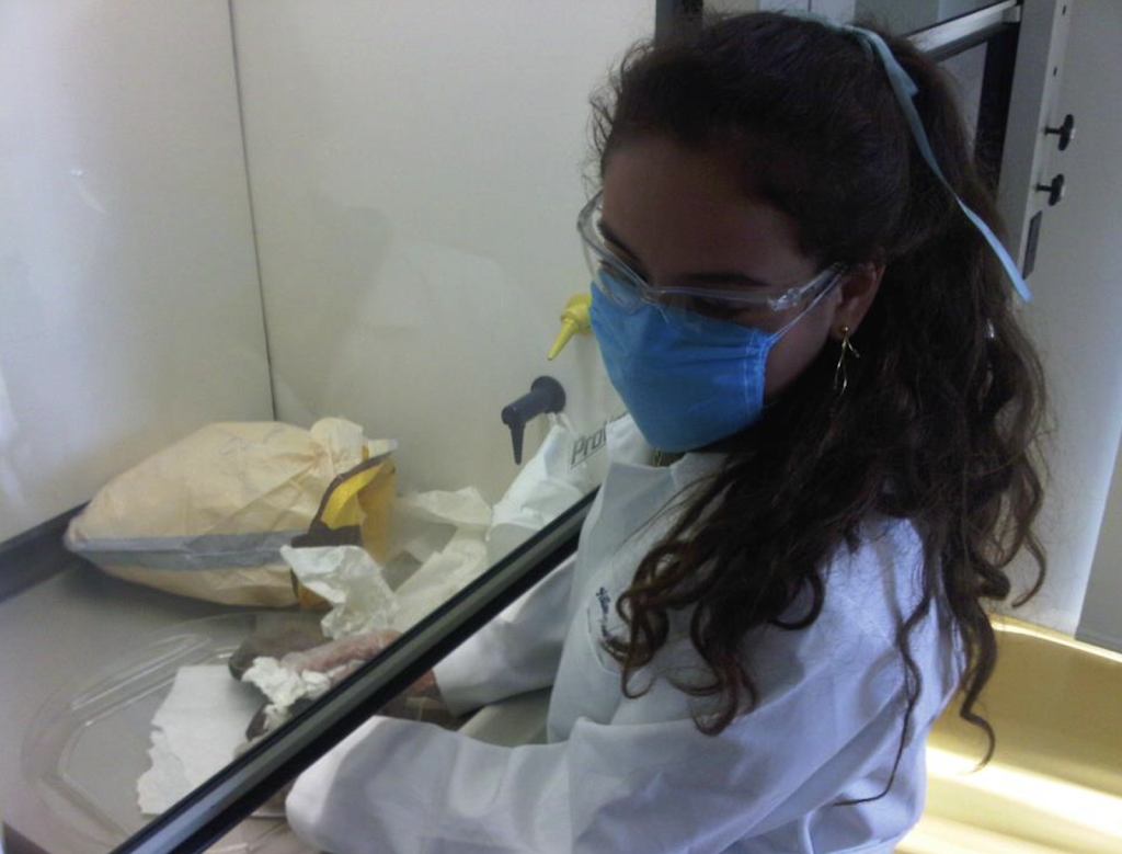
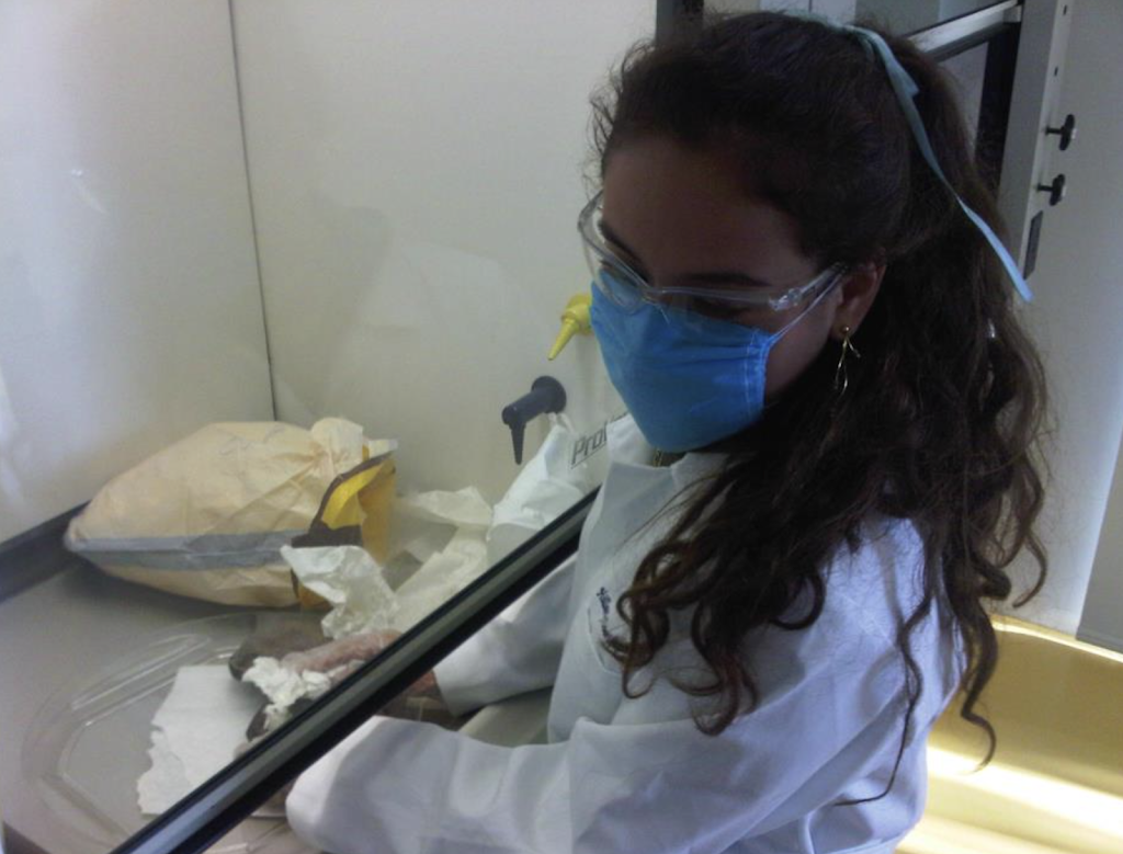
A med student at the molecular biology lab

Molecular biology is not typically a requirement for admission to medical school. However, knowledge of molecular biology can be useful for medical students, as it can provide a better understanding of the underlying mechanisms of diseases and the action of drugs at the molecular level. Medical school curriculum typically focuses on the clinical aspects of medicine, such as anatomy, physiology, and pharmacology, rather than on the molecular basis of diseases. However, some medical schools may require or recommend courses in molecular biology or other related fields, such as biochemistry, as part of their curriculum.

Molecular biology is a broad field that studies the structure, function, and behavior of molecules within living organisms. It encompasses many different sub-disciplines, including molecular genetics, genomics, proteomics, and bioinformatics. Molecular biologists use a variety of techniques, such as PCR (polymerase chain reaction), next-generation sequencing, and protein purification, to study the molecular basis of diseases and develop new treatments.

File:Guy's Hospital00.jpg

In medicine, molecular biology has many applications. For example, it can be used to identify genetic mutations that are associated with specific diseases, such as cancer or cystic fibrosis. It can also be used to develop diagnostic tests that can identify the presence of specific diseases or conditions at an early stage, when they are more treatable. Additionally, molecular biology can be used to develop new drugs or medical treatments that target specific molecules or pathways within the body.

While knowledge of molecular biology is not required for admission to medical school, it can be useful for medical students interested in gaining a deeper understanding of the underlying mechanisms of diseases and the action of drugs. Some medical schools may require or recommend courses in molecular biology as part of their curriculum, so it may be worth considering taking courses in this field if you are interested in pursuing a career in medicine.